Illustrated dissections of male genitalia
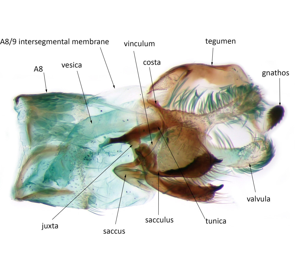
Coleophora discordella
|
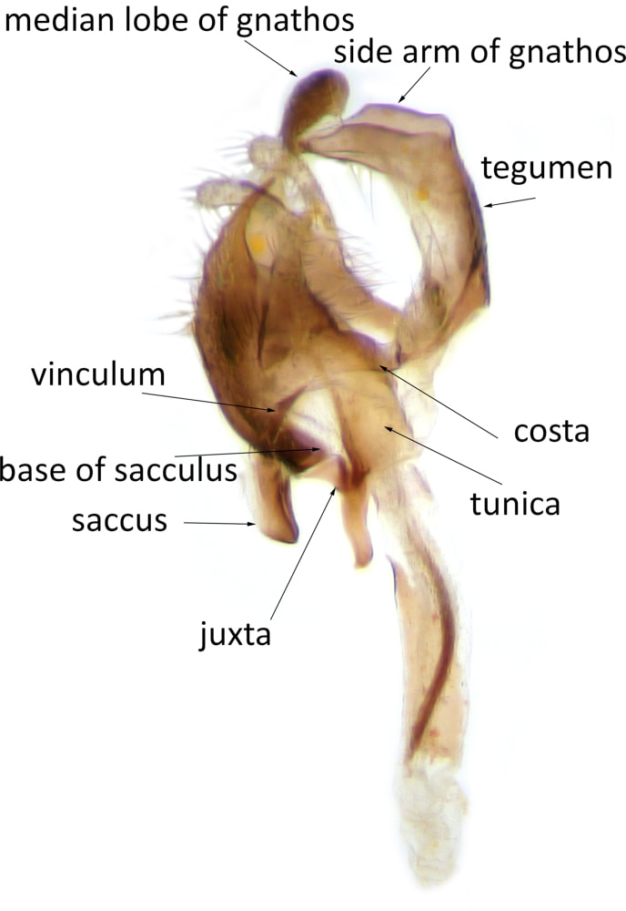
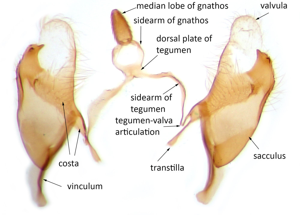
Lateral view of the genital capsule shows:
|
|
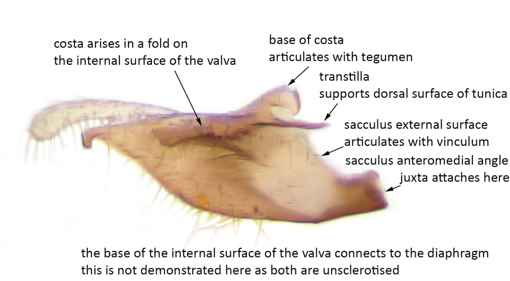
Partial medial view of the detached valva shows
- the valva has internal and external surfaces with anteroventral and posterodorsal margins
- the base of the costa divides to produce a dorsal spur that articulates with the anterior end of the tegumen and a ventral spur that extends medially as a transtilla (which in this species is very short)
- the lateral portion of the costa forms a sclerotised fold on the inner surface of the valva
- the valvula extends laterally from the lateral end of the costa and is unsclerotised and covered in short hairs
- the sacculus forms a fold on the anteroventral and lateral margins of the valva. The sclerotisation of this fold on the internal surface is fairly narrow at the base of the anteroventral margin, more extensive more distally and extends over the most of the external surface of the valva
- the internal surface of the valva between its base, the saccular and costal folds is largely hyaline.
|
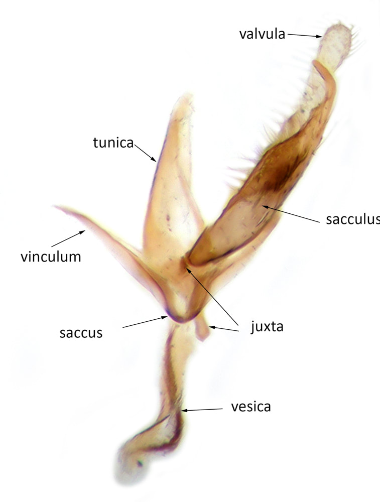
Ventral view of vinculum, valva and aedeagus shows:
|
|
Coleophora albicosta
|
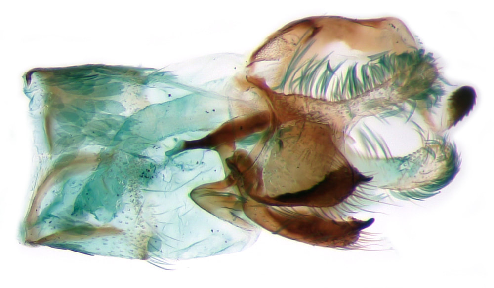
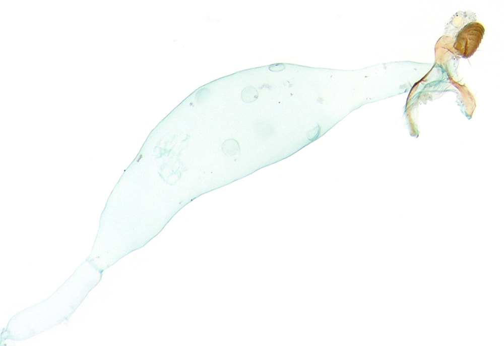
This shows a stained image of the genital capsule with the integument of A8 and A8/9 intersegmental membrane in situ. Naturally the genital capsule lies internal to A8; extension of the A8/9 intersegmental membrane (which is entirely hyaline and scale free) brings the genital capsule into a position almost entirely posterior to its attachments to the membrane. These images attempt to show these attachments - which are:
|
|
|
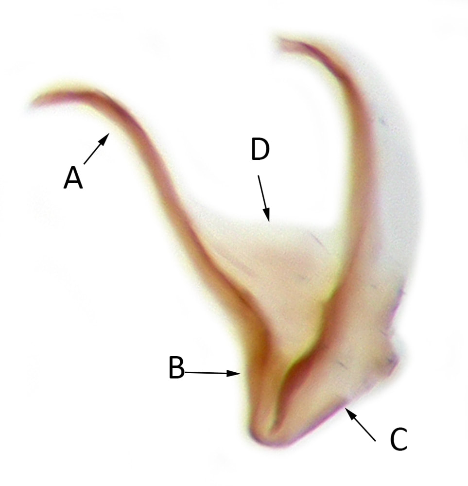
Detached vinculum (dorsal view)
|
|
Coleophora trochilella
|
Detached tegumen* (dorsal view)
Connections:
Valva and aedeagus showing connection of juxta to sacculus
|
|
Coleophora caespititiella
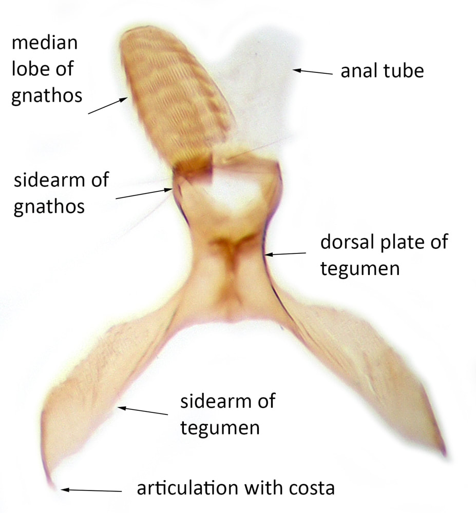
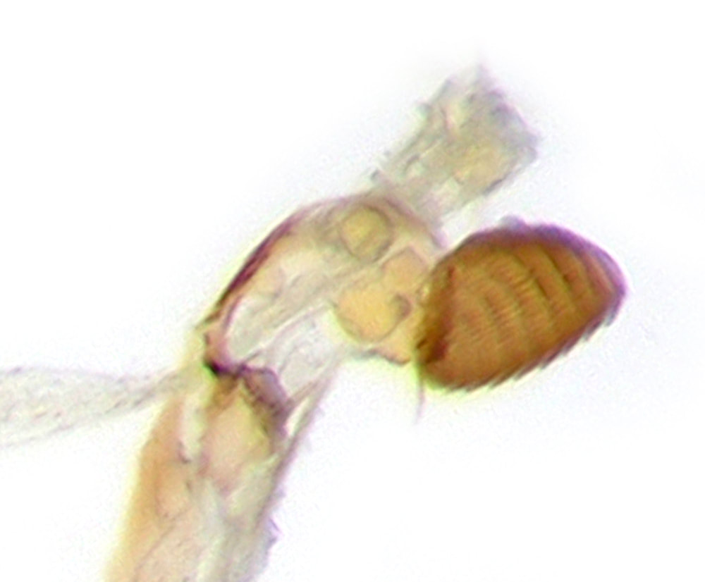
This dissection demonstrates the passage of the alimentary canal through the tegumen and the emergence of the anal canal dorsal to the median lobe of the gnathos.
Dorsal view of the genital capsule with the alimentary canal preserved. The anal canal can be seen entering the tegumen.
|
Lateral view of tegumen. The anal canal can be seen entering the tegumen from below (anterior) and passing between its dorsal and ventral surfaces and emerging between the sidearms of the gnathos dorsal to its median lobe.
Same preparation after cleaning and staining with chlorazol black
|
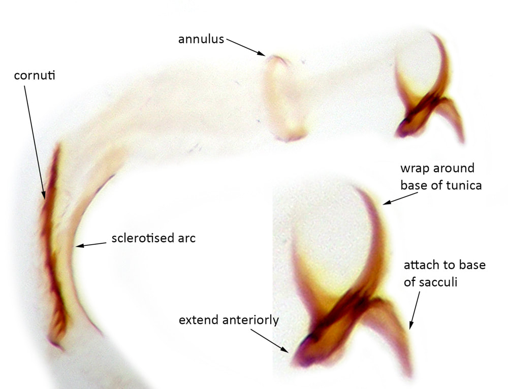
The dissection below shows the armature with the vinculum split at the apex of the saccus. It demonstrates:
- the fairly tenuous nature of the sclerotisation at the apex of the saccus, allowing easy separation of the vinculum into 2 parts
- the separation of sclerotisation at the base of the costa with an anteroventral transtilla. and a posterodorsal spur terminating at the articulation
- the fairly tenuous sclerotisation at the tegumen-valva articulation
- an extreme version of the arched type of tegumen, with a very short dorsal plate and long sidearms
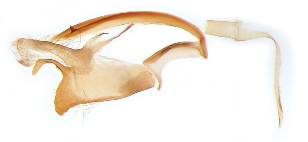
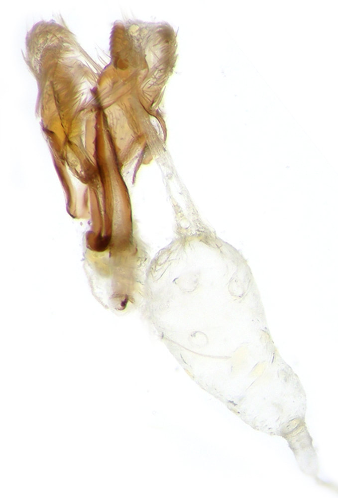
Coleophora mayrella
Usually the aedeagus should be removed with the juxta attached. Occasionally a misplaced forceps will result in detachment of the vesica from the tunica. Almost always the juxta will then remain attached to the tunica. In this dissection the juxta detached with the vesica and allows a clear demonstration of the extent of the juxta.