Female genitalia
|
|
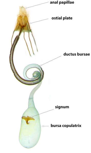
The general arrangement of the female Coleophoran genitalia is similar to that of other ditrysian Lepidoptera, and can be considered as having three main components visible in prepared specimens and used in identification of species: a posterior ovipositor; a sclerotised ring of abdominal segment 8 together with the ostium; and an anterior bursa copulatrix
1. Ovipositor
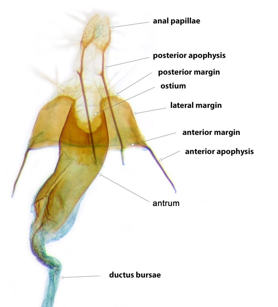
The ovipositor consists of the anal papillae, posterior apophyses and the intersegmental membrane between A9 and A8.
The anal papillae (papillae anales) surround the ovipositing and excretory orifices. They are fused anteriorly to lateral plates which bear the posterior apophyses (apophyses posteriores). The anal papillae + posterior apophyses are derived from the fused segments 9 and 10. The hyaline intersegmental membrane between A9 and A8 is infolded when the ovipositor is retracted, but allows for considerable extension when the ovipositor is protracted. The intersegmental membrane is attached posteriorly to the anterior margin of the anal papillae and anteriorly to the posterior margin of A8, such that the posterior apophyses are inside the membrane.
2. Abdominal segment 8 and the ostium
The dorsal plate of A8 ranges from being completely hyaline to completely sclerotised and fused with the ventral plate to form a complete ring. Although there are differences between species in its pattern of sclerotisation, the dorsal plate is of little interest from the identification perspective and is not usually visible on standard slide preparations with the ventral surface up. The ventral plate of A8 supports the copulatory orifice, termed the ostium, and is of critical importance in identification. The ventral plate of A8 has been termed the "subgenital plate", literally "the plate below the genitalia" - a term I consider to be nonsensical. I propose to term it the "ostial plate". Features of the ostial plate used in identification include: shape* (elongate, quadrate, transverse), position of the ostium (anterior, central, posterior), shape and extent of any incision in the posterior margin (such an incision is continuous with or leads to the ostium). **
The anterior apophyses (apophyses anteriores) project from the anterolateral margins of A8. When A8 is fully sclerotised the apophyses mark the junction between dorsal and ventral plates; when there is clear separation of dorsal and ventral plates the apophyses may be borne either at the anteroventral angles of the dorsal plate or the anterodorsal angles of the ventral plate.
3. Bursa Copulatrix
The bursa copulatrix is composed of a more-or-less discrete, narrow, tubular, posterior portion - the ductus bursae; and an ovoid or spherical, sac-like, anterior portion - the corpus bursae. In some species the most posterior part of the ductus bursae, into which the ostium opens, can be recognised as a distinct antrum***. The antrum, when present, may be strongly sclerotised, entirely or in part, and is often broader than the adjacent part of the ductus. The presence, shape and extent of the antrum and its pattern of sclerotisation are critical features used in the identification of some species. The ductus bursae may be almost entirely hyaline, but most species have an unique pattern of sclerotisation of the usually fairly straight posterior half and a hyaline coiled anterior half. Sclerotisation may be confluent and/or spiculate; it occurs in the following patterns: i) circumferential (fairly evenly distributed around the wall of the ductus), ii) hemicircumferential (fairly evenly distributed around the dorsal half of the ductus), iii) linear (a narrow line down the centre of the tube or in its wall which may continue anterior and/or posterior to other types of sclerotisation as a "tail"), iv) flanked linear (a confluent dorsal linear sclerotised ribbon with a prominent central line), v) bilinear (densely concentrated in two broad lines) vi) smudgy (poorly defined areas of sclerotisation), vii) reticulate (network pattern). It is not unusual for more than one of these patterns to be present in the same species. Coiling of the anterior half of the ductus bursae varies between species and could have some identification value, but this feature is usually not well-preserved in slide preparations.
The corpus bursae has largely hyaline walls, but usually contains a thorn-like signum, the size and shape (or absence) of which is useful in identification. A few species have a linear spiculate patch in the wall of the bursa as well as the thorn.
* The ostial plate becomes relatively more transverse with compression (eg application of a cover slip). For consistency, the assessment of shape is made on slide preparations (ie after compression) - but for the species in which this is a critical identification feature the shape is fairly obvious with or without compression.
**The length of the ostial plate is a useful tool for description of relative measurements of the apophyses and ductal sclerotisations. I have adopted the form 1xOP for example, to indicate "equal in dimension to the length of the ostial plate", ½xOP "half the length of the ostial plate" and so on.
***The antrum is synonymous with "introitus vaginae". The latter term could be deemed appropriate as it is at the opening of the copulatory canal. However, in Lepidopteran anatomy, the term vagina has been applied to the common oviduct which leads to the ovipositing pore. I think it is illogical to have the vagina and the introitus vaginae in different genital tracts and prefer to avoid both terms.
1. Ovipositor
The ovipositor consists of the anal papillae, posterior apophyses and the intersegmental membrane between A9 and A8.
The anal papillae (papillae anales) surround the ovipositing and excretory orifices. They are fused anteriorly to lateral plates which bear the posterior apophyses (apophyses posteriores). The anal papillae + posterior apophyses are derived from the fused segments 9 and 10. The hyaline intersegmental membrane between A9 and A8 is infolded when the ovipositor is retracted, but allows for considerable extension when the ovipositor is protracted. The intersegmental membrane is attached posteriorly to the anterior margin of the anal papillae and anteriorly to the posterior margin of A8, such that the posterior apophyses are inside the membrane.
2. Abdominal segment 8 and the ostium
The dorsal plate of A8 ranges from being completely hyaline to completely sclerotised and fused with the ventral plate to form a complete ring. Although there are differences between species in its pattern of sclerotisation, the dorsal plate is of little interest from the identification perspective and is not usually visible on standard slide preparations with the ventral surface up. The ventral plate of A8 supports the copulatory orifice, termed the ostium, and is of critical importance in identification. The ventral plate of A8 has been termed the "subgenital plate", literally "the plate below the genitalia" - a term I consider to be nonsensical. I propose to term it the "ostial plate". Features of the ostial plate used in identification include: shape* (elongate, quadrate, transverse), position of the ostium (anterior, central, posterior), shape and extent of any incision in the posterior margin (such an incision is continuous with or leads to the ostium). **
The anterior apophyses (apophyses anteriores) project from the anterolateral margins of A8. When A8 is fully sclerotised the apophyses mark the junction between dorsal and ventral plates; when there is clear separation of dorsal and ventral plates the apophyses may be borne either at the anteroventral angles of the dorsal plate or the anterodorsal angles of the ventral plate.
3. Bursa Copulatrix
The bursa copulatrix is composed of a more-or-less discrete, narrow, tubular, posterior portion - the ductus bursae; and an ovoid or spherical, sac-like, anterior portion - the corpus bursae. In some species the most posterior part of the ductus bursae, into which the ostium opens, can be recognised as a distinct antrum***. The antrum, when present, may be strongly sclerotised, entirely or in part, and is often broader than the adjacent part of the ductus. The presence, shape and extent of the antrum and its pattern of sclerotisation are critical features used in the identification of some species. The ductus bursae may be almost entirely hyaline, but most species have an unique pattern of sclerotisation of the usually fairly straight posterior half and a hyaline coiled anterior half. Sclerotisation may be confluent and/or spiculate; it occurs in the following patterns: i) circumferential (fairly evenly distributed around the wall of the ductus), ii) hemicircumferential (fairly evenly distributed around the dorsal half of the ductus), iii) linear (a narrow line down the centre of the tube or in its wall which may continue anterior and/or posterior to other types of sclerotisation as a "tail"), iv) flanked linear (a confluent dorsal linear sclerotised ribbon with a prominent central line), v) bilinear (densely concentrated in two broad lines) vi) smudgy (poorly defined areas of sclerotisation), vii) reticulate (network pattern). It is not unusual for more than one of these patterns to be present in the same species. Coiling of the anterior half of the ductus bursae varies between species and could have some identification value, but this feature is usually not well-preserved in slide preparations.
The corpus bursae has largely hyaline walls, but usually contains a thorn-like signum, the size and shape (or absence) of which is useful in identification. A few species have a linear spiculate patch in the wall of the bursa as well as the thorn.
* The ostial plate becomes relatively more transverse with compression (eg application of a cover slip). For consistency, the assessment of shape is made on slide preparations (ie after compression) - but for the species in which this is a critical identification feature the shape is fairly obvious with or without compression.
**The length of the ostial plate is a useful tool for description of relative measurements of the apophyses and ductal sclerotisations. I have adopted the form 1xOP for example, to indicate "equal in dimension to the length of the ostial plate", ½xOP "half the length of the ostial plate" and so on.
***The antrum is synonymous with "introitus vaginae". The latter term could be deemed appropriate as it is at the opening of the copulatory canal. However, in Lepidopteran anatomy, the term vagina has been applied to the common oviduct which leads to the ovipositing pore. I think it is illogical to have the vagina and the introitus vaginae in different genital tracts and prefer to avoid both terms.
Copulation, fertlisation and ovipositing
The functioning of the female genital tract is not very apparent from the anatomy of the genital structures that are usually preserved in a slide preparation. Although this is of little importance in a work primarily aimed at identification of the species, I feel this section would be incomplete without some explanation of functioning. At copulation the male aedeagus is inserted into the ostium and a capsule of sperm ("spermatophore') is placed in the bursa copulatrix. Sperm are released from this capsule and make their way via the ductus seminilis into the common oviduct and from there into a storage tube - the "spermatheca". The spermatheca has a divided lumen, the afferent channel transferring sperm away from the common oviduct and the efferent channel transferring sperm back to the common oviduct. Paired ovaries release eggs into an oviduct; the two oviducts unite to form the common oviduct*. Sperm fertilise eggs in the common oviduct and fertilised eggs are then released by oviposition.
In slide preparations connections between the ductus seminilis, spermatheca and common oviduct are usually destroyed during dissection. The origin of the ductus seminilis is usually discernible as a narrow tube leaving the ductus bursae at ~midway along its length, often at the anterior end of a sclerotised posterior section. The spermatheca is sometimes recognisable in part, during dissection, as a very fine very tightly coiled tube, but is seldom preserved in a slide preparation. The common oviduct may be identifiable as tube within the ovipositor, leading to the anal papillae. (see Female Dissections)
* This is the general Lepidopteran arrangement. In the few Coleophora that I have been able to identify the ovaries of, they appear to be conjoint and sessile - opening directly into the common oviduct
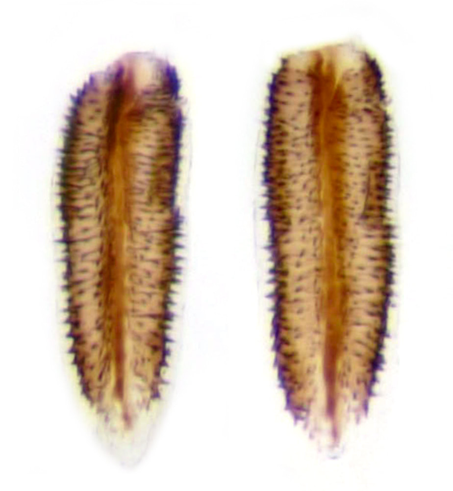
C.alticolella. Hemicircumferential, spiculate flanked linear (ventral and dorsal views)
|
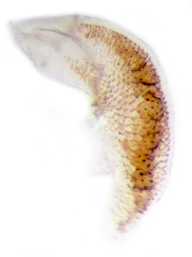
C.alticolella. Smudgy-reticulate
|
In the species descriptions I have used some shorthand notation for the important parts:
AP = Anal papillae
PA = Posterior apophyses; AA = Anterior Apophyses
A8 - Abdominal segment 8
OP = Ostial plate
PM = Posterior margin of ostial plate; AM = Anterior margin of ostial plate
BC - Bursa copulatrix
DB = Ductus bursae
CB = Corpus bursae
AP = Anal papillae
PA = Posterior apophyses; AA = Anterior Apophyses
A8 - Abdominal segment 8
OP = Ostial plate
PM = Posterior margin of ostial plate; AM = Anterior margin of ostial plate
BC - Bursa copulatrix
DB = Ductus bursae
CB = Corpus bursae
